In medical devices, visibility is essential for performing safe and effective procedures. Guiding and monitoring complex catheters and implants without adequate imaging can pose significant challenges and risks.
Medical devices with greater radiopacity are much easier for practitioners to precisely place and monitor. They’re more visible in imaging, which can make all the difference in medical procedures that demand accuracy.
At Medical Murray, we address this by innovatively incorporating radiopaque materials like barium sulfate, bismuth oxide, platinum, and gold into devices. This enables clinicians to accurately track, position, and monitor devices during surgery, thereby reducing surgical risks and enhancing patient outcomes.
Common Methods to Improve Radiopacity
Radiopaque Additive Materials
One of the most common methods to enhance radiopacity is by incorporating radiopaque materials directly into the polymer used for the device. Barium sulfate, bismuth oxide, and tungsten are popular additives that can be mixed into the polymer matrix before it is extruded or molded into form. This method ensures that the entire component extruded or molded is visible under imaging, without compromising its structural integrity.
Coatings with Radiopaque Materials
In some cases, applying a thin layer of radiopaque material to the surface of the device can enhance visibility. This method is often used when adding radiopaque materials into the polymer is not feasible or when selective areas of the device need to be highlighted.
Integrating Radiopaque Materials into Braid Reinforcement
For catheters that require a high degree of flexibility and strength, radiopaque materials can be woven into the braid reinforcement. This method allows the catheter to maintain its mechanical properties while being visible under X-ray. The braid pattern itself can be designed to optimize visibility, depending on the clinical requirements.
Incorporating Marker Bands
Marker bands are small, radiopaque rings placed at specific locations along a catheter or implant to provide clear reference points under imaging. These bands are typically made of materials like platinum, gold, or tantalum, known for their excellent radiopacity.
Seamless Marker Band Integration
Integrating marker bands into catheters requires careful consideration to ensure they do not disrupt the catheter’s performance. Here are some advanced techniques used at Medical Murray to seamlessly integrate these bands:
Encapsulation into the Catheter Wall or Tip
By embedding marker bands directly into the catheter wall during the lamination or jacketing process, we can ensure a smooth outer surface. This method involves placing the marker band between layers of the catheter’s shaft during the manufacturing process, effectively encasing it within the catheter wall. The marker band can also be insert molded into the catheter tip. These techniques provide excellent durability and ensures the bands do not interfere with the catheter’s flexibility or insertion.

Crimping and Swaging onto the Shaft
Marker bands can also be crimped and swaged directly onto the catheter shaft. This technique is straightforward and effective, allowing the bands to be securely attached without compromising the catheter’s performance. This method is particularly useful for applications where precise placement and secure attachment are critical.
Encapsulating with Subassemblies like ePTFE Wrapping
In some cases, marker bands can be encapsulated using specialized subassemblies, such as wrapping the bands with expanded polytetrafluoroethylene (ePTFE). This method offers additional protection and ensures that the marker bands are securely attached to the catheter. The ePTFE wrapping also minimizes any potential for the bands to shift or cause irritation during use.
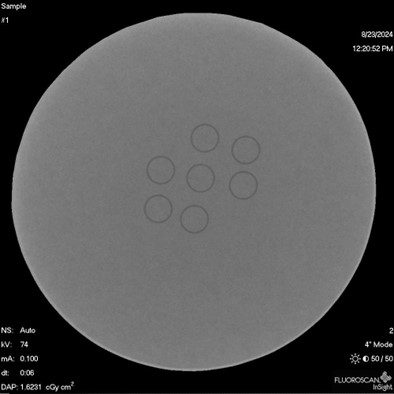
Radiopacity Testing
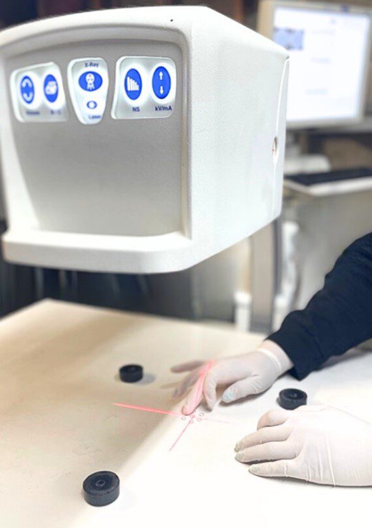
Once the radiopaque features have been integrated, testing is essential to ensure the device meets the required visibility standards. Radiopacity testing involves imaging the device under X-ray to confirm that it is visible and that the marker bands or other radiopaque elements are correctly positioned and functional. Medical Murray adheres to the ASTM F640-23 radiopacity testing standard, ensuring that all devices meet rigorous industry requirements.
At Medical Murray, we offer comprehensive radiopacity testing services as part of our development process. Our advanced imaging equipment and expertise allow us to validate the radiopacity of catheters, implants, and other devices, ensuring they perform as intended during clinical use.
Why Choose Medical Murray for Radiopacity Testing?
With years of experience in developing complex medical devices, Medical Murray is your trusted partner for enhancing radiopacity. Whether you need to incorporate radiopaque materials into your design, integrate marker bands, or validate your device’s radiopacity, our team has the expertise and capabilities to deliver. Let us help you ensure your devices are not only effective but also visible when it matters most.
Contact us today to discover how our expertise can support your next project.